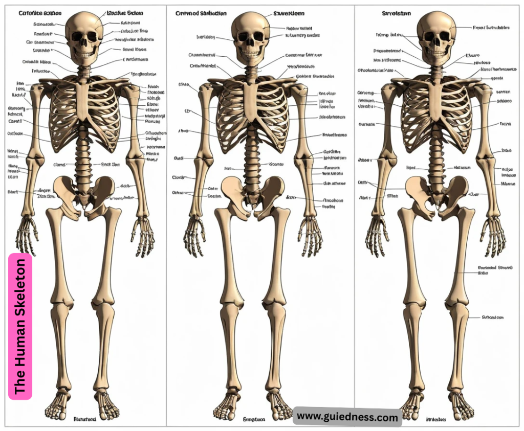
The Human Skeleton Of course. Here is a comprehensive overview of the human skeleton, covering its functions, divisions, and key facts.
The Human Skeleton: An Overview
- An adult human has 206 bones, although this number can vary slightly from person to person.
Functions of the Skeleton
- The skeleton is far more than just a structural support; it performs several critical jobs:
- Support: It provides a rigid framework that supports the body against gravity and anchors all soft organs.
- Protection: Bones protect vital internal organs. The skull protects the brain, the rib cage protects the heart and lungs, and the vertebral column protects the spinal cord.
- Movement: Bones act as levers. When skeletal muscles attached to them contract, they pull on the bones, enabling movement at the joints.
- Mineral Storage: Bones serve as a reservoir for minerals, especially calcium and phosphorus. These minerals are released into the bloodstream as needed for various bodily functions.
- Blood Cell Production: Hematopoiesis, the production of blood cells, occurs in the red bone marrow found within certain bones (e.g., pelvis, sternum, humerus, and femur).
- Fat Storage: Yellow bone marrow, found in the central cavities of long bones, stores fat (lipids) as an energy reserve.
The Axial Skeleton (80 bones)
- This forms the central axis of the body and includes the bones of the head and trunk.
Skull (28 bones + 6 ear ossicles):
- Cranium (8 bones): Frontal, 2 Parietal, 2 Temporal, Occipital, Sphenoid, Ethmoid. Protects the brain.
- Facial Bones (14 bones): Mandible (jaw), Maxilla, Zygomatic (cheekbones), Nasal, etc.
- Auditory Ossicles (6 bones): 3 tiny bones in each ear (Malleus, Incus, Stapes) that transmit sound.
- Hyoid Bone (1 bone): A U-shaped bone in the neck that supports the tongue and serves as an attachment point for muscles.
Vertebral Column (26 bones):
Cervical vertebrae (7) – the neck
- Thoracic vertebrae (12) – articulate with ribs Lumbar vertebrae (5) – lower back Sacrum (1, fused from 5) Coccyx (1, fused from 4) – tailbone
Thoracic Cage (25 bones):
Sternum (1) – breastbone
- Ribs (24, 12 pairs) – true ribs (1-7), false ribs (8-10), and floating ribs (11-12).
The Appendicular Skeleton (126 bones)
- The Human Skeleton This includes the bones of the limbs and the girdles that attach them to the axial skeleton. Pectoral (Shoulder) Girdles (4 bones):
- Clavicle (2) – collarbone Scapula (2) – shoulder blade Upper Limbs (60 bones – 30 per arm):
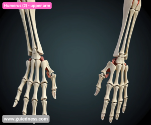
Humerus (2) – upper arm
- Radius (2) & Ulna (2) – forearm (ulna is on the pinky side) Carpals (16) – wrist bones (8 per wrist)
- Metacarpals (10) – palm bones Phalanges (28) – finger bones (14 per hand)
Pelvic (Hip) Girdle (2 bones):
- The two hip bones (ossa coxae), each formed by the fusion of the ilium, ischium, and pubis. They articulate with the sacrum at the back.
Lower Limbs (60 bones – 30 per leg):
- Femur (2) – thigh bone, the longest and strongest bone in the body
Patella (2) – kneecap
- Tibia (2) & Fibula (2) – shin bones (tibia is the larger, weight-bearing one)
- Tarsals (14) – ankle bones (7 per ankle), including the talus and the calcaneus (heel bone)
Metatarsals (10) – foot bones
Phalanges (28) – toe bones Bone Tissue and Types of Bones
- Bones are living organs made of connective tissue. They are composed of two types of osseous tissue:
- Compact Bone: The dense, hard outer layer of every bone.
- Spongy (Cancellous) Bone: The lighter, porous, honeycomb-like structure inside bones. It contains red bone marrow.
Beyond the Basics: Joints, Tissues, and Dynamic Functions
Joints (Articulations): Where Bones Meet
- Joints are classified by their structure and function, determining the range of motion between bones.
Type of Joint (by movement) Description Examples
- Synarthrosis (Immovable) Bones are tightly joined and allow no movement. Sutures in the skull.
- Amphiarthrosis (Slightly Movable) Bones are connected by cartilage and allow limited movement. Intervertebral discs between vertebrae, Pubic symphysis.
- Diarthrosis (Freely Movable) Also known as synovial joints. They have a synovial cavity filled with lubricating fluid,
- allowing for a wide range of motion. Hinge: Knee, Elbow. Ball-and-Socket: Hip, Shoulder. Pivot: Atlas/A
Structure of a Synovial Joint:
- This is the most common and complex joint type. Key structures include:
- Articular Cartilage: Smooth, slippery cartilage that covers the ends of bones, reducing friction and absorbing shock.
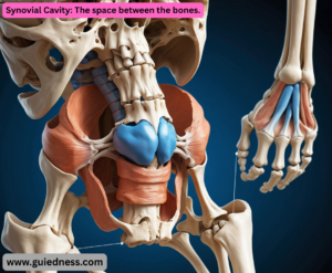
Synovial Cavity: The space between the bones.
Articular Capsule: A tough fibrous sleeve that encloses the joint.
- Ligaments, Tendons, and Bursae: Ligaments connect bone to bone, tendons connect muscle to bone, and bursae are small fluid-filled sacs that reduce friction around the joint.
The Skeleton is a Living Organ System
- Bones are not dry, static sticks. They are dynamic, living tissues with a rich blood and nerve supply.
Cellular Makeup of Bone:
- Osteoblasts: The “bone builders.” These cells synthesize and secrete bone matrix.
- Osteocytes: Mature bone cells that were once osteoblasts. They are trapped in the matrix and act as sensors for strain and damage, maintaining bone tissue.
- The Human Skeleton Osteoclasts: The “bone breakers.” These large cells dissolve and resorb bone tissue, releasing minerals into the blood. This process is crucial for bone remodeling and repair.
Bone Remodeling:
- This is a lifelong process where osteoclasts resorb old bone and osteoblasts form new bone. It serves to:
Repair micro-fractures from everyday stress.
Regulate calcium levels in the blood.
- Change the bone’s shape in response to stress (Wolff’s Law: bone grows and remodels in response to the forces placed upon it).
Common Skeletal Disorders
- Fracture: A break in a bone. Types include simple (closed), compound (open), hairline, and comminuted (shattered).
- Osteoporosis: A disease where bone density decreases, making bones porous, brittle, and highly prone to fracture. It results from an imbalance in the remodeling process (more bone is lost than formed).
- Arthritis: Inflammation of the joints. Rheumatoid Arthritis is an autoimmune disorder where the body attacks its own joint tissues.
Scoliosis: An abnormal lateral (side-to-side) curvature of the spine.
- Osteosarcoma: The most common type of bone cancer, which originates in bone-forming cells (osteoblasts).
The Skeleton Through Life
- Fetal Development: The skeleton initially consists of flexible cartilage and fibrous membranes. The process of ossification (replacing this tissue with bone) begins in the fetus and continues into early adulthood.
- Fontanelles: In newborns, the skull bones are not fully fused. The soft spots, or fontanelles, allow the skull to be compressed during birth and for rapid brain growth after.
- Growth Plates (Epiphyseal Plates): Areas of cartilage near the ends of long bones in children and adolescents. This is where bones lengthen. The plates ossify and close in early adulthood, ceasing growth in length.
- Aging: Bone mass typically peaks around age 30. After this, bone resorption slowly begins to outpace bone formation. Maintaining weight-bearing exercise and a diet rich in calcium and Vitamin D is crucial for lifelong bone health.